SEM Pathology
Table of contents
1. Examination of common tissue samples
Staining properties of platinum blue
Staining properties of osmium tetroxide
2. Examination of renal biopsy samples
3. Advantages of LVSEMs and future tasks
※Unauthorized reproduction of any materials on this website, including text and images, is strictly prohibited.
(1) Comparison of images acquired with light microscopes and with LVSEM #100002
Light microscope and LVSEM images of renal biopsy specimens
Figure No.100002a
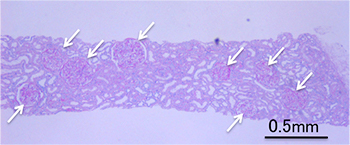
Staining: PAS staining
Microscope: Light microscope
Sample: Paraffin section (Membranous glomerulonephritis)
Magnification: ×40
Arrows: Glomeruli
Image description
Light microscope image of a renal biopsy paraffin section (membranous glomerulonephritis). Several glomeruli (→) are visible in this section.
Figure No.100002b
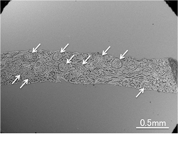
Staining: Platinum-blue staining
Microscope: LVSEM
Sample: Paraffin section (Membranous glomerulonephritis)
Accelerating voltage: 15 kV
Magnification: ×50
Arrows: Glomeruli
Image description
LVSEM image (platinum-blue staining) 5) of a paraffin section. The sections shown here and in Fig. 100002a are different, but were prepared from the same block.
Figure No.100002c
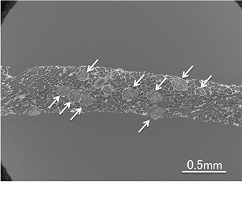
Staining: PAM staining
Microscope: LVSEM
Sample: Paraffin section (Membranous glomerulonephritis)
Accelerating voltage: 15 kV
Magnification: ×50
Arrows: Glomeruli
Image description
LVSEM image (PAM staining) of a paraffin section. The sections shown here and in Fig. 100002a are different, but were prepared from the same block.
Characteristic findings
Renal biopsy paraffin sections (mounted on glass slides) were first stained with platinum-blue or PAM, and then examined by a LVSEM. At lower magnifications (×40 - 60), all of the glomeruli (indicated by arrows) in each section can be captured as a overview image which is a similar image obtained with a light microscope. Each glomerulus is then examined at slightly higher magnifications (×600 - 1,000). Certain sliced samples show both the cross-sectional and surface views of the same glomerulus, while others provide only a large surface area of a glomerulus. At much higher magnifications (up to ×10,000), it is possible to examine areas of lesions showing pathological findings three-dimensionally.
Observations
The images shown in Figs. 100002 a, b, and c were acquired from different tissue sections, but these sections were prepared from the same paraffin block. Similar to the light microscope image (Fig. 100002a), the LVSEM images (Figs. 100002 b and c) captured at low magnifications allow for the examination of the entire section. Thus, several glomeruli (indicated by arrows) can be identified in both sections. Importantly, the light-dark patterns in images b and c are reversed. This is caused by differences in the staining specificities of platinum-blue and PAM, and provides an opportunity to observe these renal biopsy sections from complementary viewpoints.
※Unauthorized reproduction of any materials on this website, including text and images, is strictly prohibited.
