SEM Pathology
※Unauthorized reproduction of any text, image, or other content of this Website is prohibited.
A new approach to analyzing renal biopsy specimens
The 3D World Revealed by LVSEM
Nobuaki Yamanaka, M.D., Ph.D. (Director, Tokyo Renal Research Center, Professor Emeritus, Nippon Medical School)
Shinichi Okada, M.D., Ph.D. (Associate Professor Division of Pediatrics and Perinatology, Faculty of Medicine, Tottori University)
Sumire Inaga, Ph.D. (Junior Associate Professor, Department of Anatomy, Faculty of Medicine, Tottori University)
Using the distinctive characteristics of the low-vacuum scanning electron microscope (LVSEM), Dr. Sumire Inaga of the Department of Anatomy at the Faculty of Medicine of Tottori University developed a seminal new method for renal biopsy histopathologic analysis. The method represents a departure from the conventional perspective underlying the commonly applied transmission electron microscopy (TEM).
In this method, paraffin sections are prepared for light-microscope observation in three dimensions at the electronic microscopic level using the LVSEM.
Today, we invited its developer, Dr. Inaga, together with Dr. Nobuaki Yamanaka, an expert in renal pathology and nephropathy diagnosis and Director of the Tokyo Renal Research Center and Dr. Shinichi Okada, an Associate Professor in the Division of Pediatrics and Perinatology at the Faculty of Medicine of Tottori University, who has used the LVSEM to perform histopathologic analysis in collaboration with Dr. Inaga, to join us at the Tokyo Solutions Laboratory of Hitachi High-Tech Corporation todiscuss the context and salience of the development by Dr. Inaga, as well as its future potential.
1. A new approach for analyzing renal biopsy specimens using the LVSEM
The subjective symptoms of renal diseases do not emerge until the disease has reached an advanced stage, and they are generally regarded as difficult to classify. The structure and functions of the kidney are highly complex, and biopsies are particularly important for determining the relationship between disease progression and the morphology of the lesions involved. In the renal biopsy method developed in 1951, a small portion of renal tissue is excised using a slender needle and observed under a light microscope. This observation technique has since been complemented by renal immunohistopathology, in which information on immunologic reactions is obtained based on the presence and patterns of immunoglobulins, complementcomponents, and other factors, and by electron-microscopic analysis. Today the information from all three sources is combined to distinguish diseases, determine the prognosis, and choose the appropriate therapy, and these special examinations are now performed routinely in renal biopsy analysis.
The use of electron microscopy in renal biopsy analysis has generated many new information. Most electron microscopic observations are performed using TEM, which requires a high level of knowledge and sophisticated techniques for sample preparation and image analysis as well as several weeks to obtain a defi nitive diagnosis.
Dr. Inaga’s work in the Department of Anatomy at the Faculty of Medicine of Tottori University led her to wonder whether TEM, with these requirements, could really meet the needs of clinicians. This led to her desire for a simpler, faster method for conducting renal biopsy histopathologic analysis and ultimately to her development of the new method using the LVSEM. She had the opportunity to prepare biological samples for LVSEM for Dr. Kei-ichi Tanaka, who is a worldleading authority on SEM and the former professor of the lab. For, after his retirement from Tottori University, he changed the analytic tool from the ultra-high-resolution SEM with highvacuum conditions to the low-vacuum SEM with backscatter electron (BSE) mode. LVSEM cannot reach TEM in terms of resolution, but enables the observation of samples that contain oils or water or are nonconductive. In addition, the LVSEM is relatively simple to operate and requires no special sample processing.
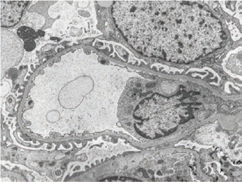
TEM images of renal biopsy specimens from cases of Alport syndrome (left) and thin basement membrane disease (right) are shown here for comparison of typical TEM images of these two pathologies.
※Unauthorized reproduction of any text, image, or other content of this Website is prohibited.
