3. Electron microscope–level analysis using paraffin sections
On his first encounter with renal histopathologic analysis by LVSEM, Dr. Nobuaki Yamanaka, a leading expert in renal pathology and nephropathy diagnosis and Director of the Tokyo Renal Research Center, intuitively understood its importance as a new methodology and began a series of investigations into its potential uses.
He soon discovered that applying the low-vacuum characteristics of LVSEM to the specimen by placing it on a slide glass in the sample chamber and observe it through the electron microscope made it possible to observe ordinary, lightmicroscope samples at the electron-microscope level, without the need for additional processing.
The combination of platinum blue with silver staining by PAM would heighten the analytical effectiveness of this method. PAM is well-established stain for analyzing renal biopsy specimens under a light microscope. As described by Dr. Inaga, “PAM stain is particularly advantageous for beautifully showing the component structure in the renal basement membrane. I noted that it was effectively the opposite of platinum-blue staining and realized these two stains could be used to obtain mutually complementary effects for observation.”
She continues, “In particular, PAM staining has a long history, and Dr. Yajima, the former professor of Dr. Yamanaka’s lab., improved PAM staining in Japan, which led to its present remarkable ease of use. PAM staining and platinumblue staining are an excellent match, which is particularly advantageous for diagnosis of renal pathologies. I think the ability to see things with one that cannot be seen with the other and to use two specimens taken from the same tissue for comparative observation can bring greater depth to diagnosis.”
Dr. Okada of the Faculty of Medicine at Tottori University, has worked in collaboration with Dr. Inaga in renal histopathology analysis, and in the perspective of the clinician on future possibilities for application of the LVSEM analysis method, he notes that “With an LVSEM, you can observe the renal biopsy tissue both at high magnifi cation and in its entirety from corner to corner, as in a light microscope. That’s an important advantage.”
“In renal histological disease, lesions occur uniformly throughout the organ in most cases, but in focal segmental glomerulosclerosis (FSGS) and some other cases, the lesion initially occurs in one part before spreading to the entire organ. If you can’t fi nd that part and see the lesion, you can’t recognize the abnormality.”
“When using TEM, if an abnormality happens to be located outside the small part of TEM specimen under observation, then the diagnosis tends to be ‘free from abnormality’. But you don’t really know whether abnormality is absent from the entire tissue. I think the capability of an LVSEM for observation of a wide area is very useful in examining for kidney pathologies in which the lesion begins in one part of the organ.”

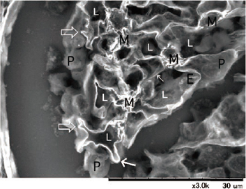
LVSEM images of a glomerulus in paraffin sections from a case diagnosed as FSGS, showing surface stained with platinum blue (left) and a cross-section stained with PAM (right).